Retinal Detachment
Learn about retinal detachment, including warning signs, causes, diagnosis, and emergency treatment options to help protect vision.
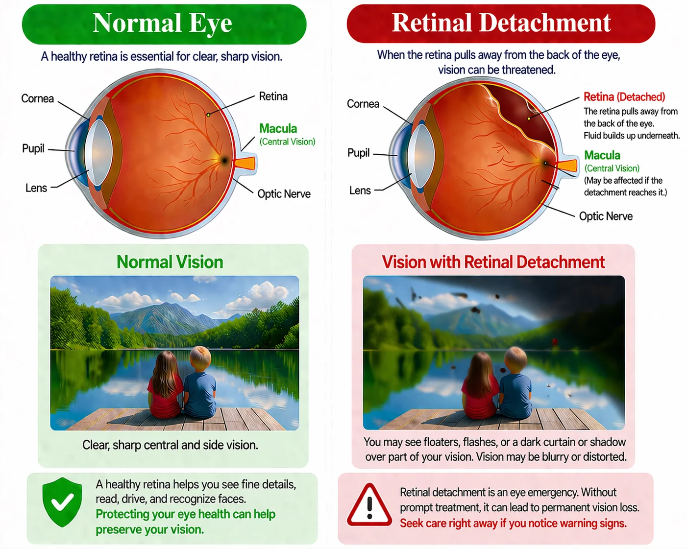
Overview
Retinal detachment is a serious eye emergency that happens when the retina pulls away from the layer beneath it that provides oxygen and nourishment. Because the retina is responsible for capturing light and sending visual signals to the brain, a detachment can lead to permanent vision loss if it is not treated quickly. Some detachments develop after a retinal tear allows fluid to move underneath the retina, while others may happen because of scar tissue or fluid buildup from other eye conditions. Early diagnosis and prompt treatment are critical to protecting as much vision as possible.
Learn more about Retinal Detachment
Do I have Retinal Detachment ?
Retinal detachment is usually painless, but the warning signs often appear before vision is permanently affected.
Symptoms of Retinal Detachment
Symptoms can include, but are not limited to:
Sudden flashes and floaters
A rapid increase in floaters or flashes of light can be an important warning sign.
Blurred or reduced side vision
Vision may suddenly become blurry or seem narrower at the edges.
A curtain, shade, or shadow over vision
A dark shadow moving across part of the visual field is an emergency warning sign.
Treatment of Retinal Detachment
Treatment depends on whether there is a tear, a limited detachment, or a more advanced detachment:
Laser or cryopexy
If a tear is found before a full detachment develops, sealing it early may prevent worse damage.
Pneumatic retinopexy
A gas bubble may be used in selected cases to press the retina back into place.
Scleral buckle or vitrectomy
More extensive detachments often require surgery to relieve traction and reattach the retina.
Whether you need a routine eye exam, upadet glasses or contacts, or help with an eye concert, our fiendly team is here to make the process easy.
- Personalized Care
- States-f-the-Art Equipment
- Experienced Optometrists
- Family-Friendly Environment
What causes Retinal Detachment?
Retinal detachment can happen in several different ways. The most common type starts with a retinal tear or hole that allows fluid to collect under the retina and lift it away from the back of the eye. This is often related to age-related changes in the vitreous. Other detachments can happen when scar tissue pulls on the retina or when fluid collects underneath it without a tear. Risk may be higher in people who are very nearsighted, have had previous cataract surgery or eye trauma, have another retinal problem, or have a family history of retinal detachment.
Getting a Diagnosis of Retinal Detachment
Retinal detachment is diagnosed during a prompt retinal evaluation. Because time matters, sudden symptoms should be checked right away, even if they seem to come and go.
Common tests used to diagnose retinal detachment include:
Dilated Eye Exam
Eye drops are used to widen the pupil so your doctor can closely inspect the retina for holes, tears, or detachment.
Ultrasound of the Eye
If bleeding or another problem makes the retina hard to see directly, ultrasound may help your doctor evaluate the inside of the eye.
Retinal Examination of Both Eyes
Your doctor may carefully examine both eyes, even if symptoms are only happening in one, because the other eye can also have retinal risk factors or tears.
Different types of Retinal Detachment
Retinal detachment is usually divided into types based on what is pulling or lifting the retina away from the back of the eye.
- Rhegmatogenous detachment is the most common type and begins with a retinal hole or tear that allows fluid to move underneath the retina;
- Tractional detachment happens when scar tissue pulls on the retina, which is often seen in people with poorly controlled diabetes;
- Exudative detachment occurs when fluid builds up under the retina without a tear and may be related to inflammation, tumors, injury, or other disease.
Factors Increasing Risk of Retinal Detachment
Some people are more likely to develop a retinal detachment than others, especially if they already have retinal stress or a history of eye problems.
Common risk factors for retinal detachment include:
- Age over 50
- Extreme nearsightedness
- Previous retinal detachment in one eye
- Family history of retinal detachment
- Previous cataract surgery or other eye surgery
- Severe eye injury or trauma
- Another retinal or eye condition
Questions about Retinal Detachment?
We’re here to help you act quickly when sudden retinal symptoms appear. Whether you are seeing new flashes and floaters, a curtain over your vision, or have already been told you have a retinal tear or detachment, our team is happy to help guide you toward prompt evaluation and treatment.
Retinal detachment is an emergency condition in which the retina pulls away from the tissue that supports and nourishes it. Without quick treatment, it can lead to permanent vision loss.
Warning signs can include a sudden increase in floaters, flashes of light, blurred vision, reduced side vision, and a curtain or shadow moving across part of your vision. These symptoms should be evaluated right away.
Treatment depends on the type and severity of the problem and may include laser therapy, cryopexy, pneumatic retinopexy, scleral buckle surgery, vitrectomy, or a combination of treatments.
Immediately. Retinal detachment is a medical emergency, and even symptoms that seem mild should be checked promptly because early diagnosis and treatment give the best chance of protecting vision.